SPTBN1
Protein-coding gene in the species Homo sapiens
| SPTBN1 | |||||||||||||||||||||||||||||||||||||||||||||||||||
|---|---|---|---|---|---|---|---|---|---|---|---|---|---|---|---|---|---|---|---|---|---|---|---|---|---|---|---|---|---|---|---|---|---|---|---|---|---|---|---|---|---|---|---|---|---|---|---|---|---|---|---|
 | |||||||||||||||||||||||||||||||||||||||||||||||||||
| Identifiers | |||||||||||||||||||||||||||||||||||||||||||||||||||
| Aliases | SPTBN1, ELF, HEL102, SPTB2, betaSpII, spectrin beta, non-erythrocytic 1, DDISBA | ||||||||||||||||||||||||||||||||||||||||||||||||||
| External IDs | OMIM: 182790; MGI: 98388; HomoloGene: 2354; GeneCards: SPTBN1; OMA:SPTBN1 - orthologs | ||||||||||||||||||||||||||||||||||||||||||||||||||
| |||||||||||||||||||||||||||||||||||||||||||||||||||
| |||||||||||||||||||||||||||||||||||||||||||||||||||
| |||||||||||||||||||||||||||||||||||||||||||||||||||
| |||||||||||||||||||||||||||||||||||||||||||||||||||
| |||||||||||||||||||||||||||||||||||||||||||||||||||
| Wikidata | |||||||||||||||||||||||||||||||||||||||||||||||||||
| |||||||||||||||||||||||||||||||||||||||||||||||||||


Spectrin beta chain, brain 1 is a protein that in humans is encoded by the SPTBN1 gene.[5]
Function
Spectrin is an actin crosslinking and molecular scaffold protein that links the plasma membrane to the actin cytoskeleton, and functions in the determination of cell shape, arrangement of transmembrane proteins, and organization of organelles. It is composed of two antiparallel dimers of alpha- and beta- subunits. This gene is one member of a family of beta-spectrin genes. The encoded protein contains an N-terminal actin-binding domain, and 17 spectrin repeats that are involved in dimer formation. Multiple transcript variants encoding different isoforms have been found for this gene.[5]
Interactions
SPTBN1 has been shown to interact with Merlin.[6]
References
- ^ a b c GRCh38: Ensembl release 89: ENSG00000115306 – Ensembl, May 2017
- ^ a b c GRCm38: Ensembl release 89: ENSMUSG00000020315 – Ensembl, May 2017
- ^ "Human PubMed Reference:". National Center for Biotechnology Information, U.S. National Library of Medicine.
- ^ "Mouse PubMed Reference:". National Center for Biotechnology Information, U.S. National Library of Medicine.
- ^ a b "Entrez Gene: SPTBN1 spectrin, beta, non-erythrocytic 1".
- ^ Neill GW, Crompton MR (September 2001). "Binding of the merlin-I product of the neurofibromatosis type 2 tumour suppressor gene to a novel site in beta-fodrin is regulated by association between merlin domains". Biochem. J. 358 (Pt 3): 727–35. doi:10.1042/0264-6021:3580727. PMC 1222106. PMID 11535133.
Further reading
- Hu RJ, Watanabe M, Bennett V (1992). "Characterization of human brain cDNA encoding the general isoform of beta-spectrin". J. Biol. Chem. 267 (26): 18715–22. doi:10.1016/S0021-9258(19)37020-6. PMID 1527002.
- Yoon SH, Skalka H, Prchal JT (1989). "Presence of erythroid and nonerythroid spectrin transcripts in human lens and cerebellum". Invest. Ophthalmol. Vis. Sci. 30 (8): 1860–6. PMID 2474519.
- Chang JG, Scarpa A, Eddy RL, Byers MG, Harris AS, Morrow JS, Watkins P, Shows TB, Forget BG (1993). "Cloning of a portion of the chromosomal gene and cDNA for human beta-fodrin, the nonerythroid form of beta-spectrin". Genomics. 17 (2): 287–93. doi:10.1006/geno.1993.1323. PMID 8406479.
- Shimizu T, Takakuwa Y, Koizumi H, Ishibashi T, Ohkawara A (1996). "Calcium-dependent peripheral localization of 4.1-like proteins and fodrin in cultured human keratinocytes". Biol. Cell. 86 (1): 19–26. doi:10.1016/0248-4900(96)89520-7. PMID 8688828.
- Bonaldo MF, Lennon G, Soares MB (1997). "Normalization and subtraction: two approaches to facilitate gene discovery". Genome Res. 6 (9): 791–806. doi:10.1101/gr.6.9.791. PMID 8889548.
- Holleran EA, Tokito MK, Karki S, Holzbaur EL (1997). "Centractin (ARP1) associates with spectrin revealing a potential mechanism to link dynactin to intracellular organelles". J. Cell Biol. 135 (6 Pt 2): 1815–29. doi:10.1083/jcb.135.6.1815. PMC 2133946. PMID 8991093.
- Djinovic Carugo K, Bañuelos S, Saraste M (1997). "Crystal structure of a calponin homology domain". Nat. Struct. Biol. 4 (3): 175–9. doi:10.1038/nsb0397-175. PMID 9164454. S2CID 1428195.
- Scoles DR, Huynh DP, Morcos PA, Coulsell ER, Robinson NG, Tamanoi F, Pulst SM (1998). "Neurofibromatosis 2 tumour suppressor schwannomin interacts with betaII-spectrin". Nat. Genet. 18 (4): 354–9. doi:10.1038/ng0498-354. PMID 9537418. S2CID 30709629.
- Sihag RK (1998). "Brain beta-spectrin phosphorylation: phosphate analysis and identification of threonine-347 as a heparin-sensitive protein kinase phosphorylation site". J. Neurochem. 71 (5): 2220–8. doi:10.1046/j.1471-4159.1998.71052220.x. PMID 9798950. S2CID 23703252.
- Bañuelos S, Saraste M, Djinović Carugo K (1999). "Structural comparisons of calponin homology domains: implications for actin binding". Structure. 6 (11): 1419–31. doi:10.1016/S0969-2126(98)00141-5. PMID 9817844.
- Löfvenberg L, Backman L (1999). "Calpain-induced proteolysis of beta-spectrins". FEBS Lett. 443 (2): 89–92. doi:10.1016/S0014-5793(98)01697-4. PMID 9989581. S2CID 85055199.
- Hayes NV, Scott C, Heerkens E, Ohanian V, Maggs AM, Pinder JC, Kordeli E, Baines AJ (2000). "Identification of a novel C-terminal variant of beta II spectrin: two isoforms of beta II spectrin have distinct intracellular locations and activities". J. Cell Sci. 113 (11): 2023–34. doi:10.1242/jcs.113.11.2023. PMID 10806113.
- Kontrogianni-Konstantopoulos A, Frye CS, Benz EJ, Huang SC (2001). "The prototypical 4.1R-10-kDa domain and the 4.1g-10-kDa paralog mediate fodrin-actin complex formation". J. Biol. Chem. 276 (23): 20679–87. doi:10.1074/jbc.M010581200. PMID 11274145.
- Neill GW, Crompton MR (2001). "Binding of the merlin-I product of the neurofibromatosis type 2 tumour suppressor gene to a novel site in beta-fodrin is regulated by association between merlin domains". Biochem. J. 358 (Pt 3): 727–35. doi:10.1042/0264-6021:3580727. PMC 1222106. PMID 11535133.
- Chen Y, Yu P, Lu D, Tagle DA, Cai T (2002). "A novel isoform of beta-spectrin II localizes to cerebellar Purkinje-cell bodies and interacts with neurofibromatosis type 2 gene product schwannomin". J. Mol. Neurosci. 17 (1): 59–70. doi:10.1385/JMN:17:1:59. PMID 11665863. S2CID 6524087.
- Shoeman RL, Hartig R, Hauses C, Traub P (2003). "Organization of focal adhesion plaques is disrupted by action of the HIV-1 protease". Cell Biol. Int. 26 (6): 529–39. doi:10.1006/cbir.2002.0895. PMID 12119179. S2CID 39778155.
- Tomsig JL, Snyder SL, Creutz CE (2003). "Identification of targets for calcium signaling through the copine family of proteins. Characterization of a coiled-coil copine-binding motif". J. Biol. Chem. 278 (12): 10048–54. doi:10.1074/jbc.M212632200. PMID 12522145.
- Tang Y, Katuri V, Dillner A, Mishra B, Deng CX, Mishra L (2003). "Disruption of transforming growth factor-beta signaling in ELF beta-spectrin-deficient mice". Science. 299 (5606): 574–7. Bibcode:2003Sci...299..574T. doi:10.1126/science.1075994. PMID 12543979. S2CID 83664290.
- Robb VA, Li W, Gascard P, Perry A, Mohandas N, Gutmann DH (2003). "Identification of a third Protein 4.1 tumor suppressor, Protein 4.1R, in meningioma pathogenesis" (PDF). Neurobiol. Dis. 13 (3): 191–202. doi:10.1016/S0969-9961(03)00071-8. PMID 12901833. S2CID 46574223.
- v
- t
- e
PDB gallery
-
 1aa2: CALPONIN HOMOLOGY (CH) DOMAIN FROM HUMAN BETA-SPECTRIN
 -
 1bkr: CALPONIN HOMOLOGY (CH) DOMAIN FROM HUMAN BETA-SPECTRIN AT 1.1 ANGSTROM RESOLUTION
 -
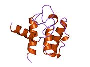
 1btn: STRUCTURE OF THE BINDING SITE FOR INOSITOL PHOSPHATES IN A PH DOMAIN
 -
 1mph: PLECKSTRIN HOMOLOGY DOMAIN FROM MOUSE BETA-SPECTRIN, NMR, 50 STRUCTURES


