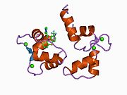
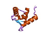
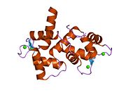
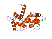
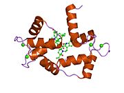
Calmodulin 1
Protein-coding gene in the species Homo sapiens
| CALM1 | |||||||||||||||||||||||||||||||||||||||||||||||||||
|---|---|---|---|---|---|---|---|---|---|---|---|---|---|---|---|---|---|---|---|---|---|---|---|---|---|---|---|---|---|---|---|---|---|---|---|---|---|---|---|---|---|---|---|---|---|---|---|---|---|---|---|
 | |||||||||||||||||||||||||||||||||||||||||||||||||||
| Identifiers | |||||||||||||||||||||||||||||||||||||||||||||||||||
| Aliases | CALM1, CALML2, CAMI, CPVT4, DD132, PHKD, caM, LQT14, Calmodulin 1, calmodulin 1 (phosphorylase kinase, delta), CAMC, CAMB, CAMIII, CAM3, CAM2 | ||||||||||||||||||||||||||||||||||||||||||||||||||
| External IDs | OMIM: 114180; HomoloGene: 134804; GeneCards: CALM1; OMA:CALM1 - orthologs | ||||||||||||||||||||||||||||||||||||||||||||||||||
| |||||||||||||||||||||||||||||||||||||||||||||||||||
| |||||||||||||||||||||||||||||||||||||||||||||||||||
| |||||||||||||||||||||||||||||||||||||||||||||||||||
| |||||||||||||||||||||||||||||||||||||||||||||||||||
| Wikidata | |||||||||||||||||||||||||||||||||||||||||||||||||||
| |||||||||||||||||||||||||||||||||||||||||||||||||||


Calmodulin 1 is a protein in humans that is encoded by the CALM1 gene.[3]
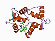
Calmodulin[4] plays a role in calcium signal transduction pathways by regulating control of ion channels, enzymes, aquaporins, and other proteins. It functions as a calcium-binding protein that has been grouped into the EF-hand motif found in eukaryotic cells. Calmodulin plays a significant role in numerous cellular pathways and it acts as a calcium detector within the cells that interact with varied target proteins. Additionally, it simulates[5] the activation of over twenty amino acids which helps to control various physiological functions. It is also required for various regulatory roles in cell proliferation and throughout many points during the cell cycle.
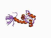
Upon binding to targeted calcium (acts as ligand), calmodulin undergoes a change in shape that allows it to interact with multiple protein types including phosphatases, ion channels, and kinases. This conformational change is associated with undergoing various cellular processes: including muscle contraction, release of neurotransmitters into the bloodstream, and gene expression.
Function
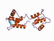
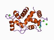
Calmodulin 1 is the archetype of the family of calcium-modulated (calmodulin) proteins of which nearly 20 members have been found. They are identified by their occurrence in the cytosol or on membranes facing the cytosol and by a high affinity for calcium. Calmodulin contains 149 amino acids and has 4 calcium-binding EF hand motifs. Its functions include roles in growth and the cell cycle as well as in signal transduction and the synthesis and release of neurotransmitters.[6]
Gene expression
In humans, there are three genetic isoforms of calmodulin which are encoded by the homologous gene variations: CALM1, CALM2, and CALM3. Each three of the isoforms produce distinct, yet closely associated forms of calmodulin. At the nucleic acid level, the coding regions differ by a 15% between CALM1 and CALM2 and 13% between CALM2 and CALM3.[7]
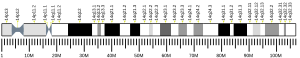
Calmodulin I, abbreviated CALM1, is located on chromosome 14 of the human genome, and is one of the three isoforms of calmodulin. It’s found in all human tissues, although the expression varies depending on tissue type. There are high expression levels found in the brain, muscle, and blood.
Throughout the body, CALM1 plays a significant role in muscle contraction and relaxation in skeletal and smooth muscle. In heart muscle, CALM1 is vital for the regulation of calcium signaling to control efficient cardiac functioning. Calcium/calmodulin protein kinases (CaMKs)[8] work symbiotically to regulate calcium signaling throughout the body. CAMKII, the most prolific isoform, is found in cardiac tissue where it controls excitation-contraction coupling. Calmodulin I also plays an important role in the immune system through lymphocytes (white blood cells) where it contributes to immune cell function and activation. In bone tissue, Calmodulin I is associated with osteoblasts, osteoclasts, and osteocytes, by functioning in intracellular calcium signaling to ensure bone mineralization, resorption, and remodeling.
Calmodulin 1[7] can be expressed as one of two transcript types, which can be distinguished by length and tissue location. The major transcript is present in all tissues and is recorded as 1.7-kb in length. The minor transcript is either 4.1-kb or 4.4kb in length, and is only found in brain and skeletal muscle tissue. The difference in transcript lengths are caused by substitute cleavage and polyadenylation signals (APA), which permits the origination of different mRNA isoforms.
Pseudogenes
There are two known pseudogenes of Calmodulin 1, which are known as CALMIPI and CALMIP2. CALMPI was first discovered on chromosome 7, and the CALMPI2 was later identified on chromosome X. Experimentation shows both pseudogenes lack introns and have multiple mutations in their open reading frame, meaning that they cease all functions.
Mapping
Human[9] and rodent hybridized somatic cell panels show that complementary DNA for calmodulin I was localized to chromosome 14, with some cross hybridization activity on chromosome 7, and minor involvement on chromosome X.
Identifiers
- Protein Identification: P62158 (aka CALM_HUMAN)
- Gene Identification: 114180
| CALM1 Biochemical & Signaling Pathways[10] Kyoto Encyclopedia of Genes and Genomes (KEGG):[11] | |
|---|---|
| hsa04020 | Calcium signaling pathway |
| hsa04070 | Phosphatidylinositol signaling system |
| hsa04114 | Oocyte meiosis |
| hsa04270 | Vascular smooth muscle contraction |
| hsa04720 | Long-term potentiation |
| hsa04722 | Neurotrophin signaling pathway |
| hsa04740 | Olfactory transduction |
| hsa04744 | Phototransduction |
| hsa04910 | Insulin signaling pathway |
| hsa04912 | GnRH signaling pathway |
| hsa04916 | Melanogenesi |
| hsa05010 | Alzheimer's disease |
| hsa05214 | Glioma |
| Calmodulin I protein family domains: | |
|---|---|
| PF00036 | EF hand |
| PF08726 | Ca2+ insensitive EF hand |
| PF12763 | Cytoskeletal-regulatory complex EF hand |
| PF13202 | EF hand |
| PF13405 | EF-hand domain |
| PF13499 | EF-hand domain pair |
| PF13833 | EF-hand domain pair |
| PF14658 | EF-hand domain |
Interactions
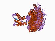
Calmodulin 1 has been shown to interact with:
Mutations
Mutations of CALM1 CALM2 or CALM3 can lead to critical cardiac deficiencies including long QT syndrome (LQTS) and catecholaminergic polymorphic ventricular tachycardia (CPVT).[18] Studies investigating calmodulin-associated diseases have discovered multiple proteins modified by calmodulin that determine the virulence of the mutations, including the cardiac L-type calcium channel (LTCC) Cav1.2, the sarcoplasmic reticulum calcium release channel, and the ryanodine receptor 2 (RyR2).
CALM1 disease mutations are often diagnosed in patients aged ten or younger, whereas CALM2 and CALM3 mutations typically develop in adulthood. Calmodulin functioning defects cause interference of vital calcium signaling events within the heart muscle which disrupts membrane ion channels. The disruptions in cell signaling can lead to potentially life-threatening cardiac disturbances in adolescence.
- Long QT Syndrome-14 (LQT14)
LQT14[19] is caused by the heterozygous mutation in the CALM1 gene (114180) on chromosome 14q32. It often produces life-threatening ventricular arrhythmias that manifest at a young age with persistent periods of T-wave alternans, notably sustained QTc intervals, and irregular 2:1 atrioventricular blocks.
CPVT[20] is an inherited disorder that presents with episodes of syncope and/or sudden cardiac infarctions during exercise or extreme emotional episodes in humans without structural cardiac deformities. Mutations in the ryanodine-receptor 2 channel (RYR2) that causes calcium leakage from the sarcoplasmic reticulum have been proven to cause about half of dominantly inherited cases of CPVT.
It has been discovered that some individuals with CPVT have distinct mutations on the Calmodulin I gene. The mutations cause disruption in the proper functioning of the gene, which leads to abnormal calcium control in cardiac tissue cells. The calcium disturbance can trigger ventricular arrhythmias in reaction to blood vessel vasoconstriction, such as during periods of exercise or elevated stress.
References
- ^ a b c GRCh38: Ensembl release 89: ENSG00000198668 – Ensembl, May 2017
- ^ "Human PubMed Reference:". National Center for Biotechnology Information, U.S. National Library of Medicine.
- ^ Wawrzynczak EJ, Perham RN (August 1984). "Isolation and nucleotide sequence of a cDNA encoding human calmodulin". Biochemistry International. 9 (2): 177–185. PMID 6385987.
- ^ "UniProt". www.uniprot.org. Retrieved 2023-10-11.
- ^ Means AR, VanBerkum MF, Bagchi I, Lu KP, Rasmussen CD (1991). "Regulatory functions of calmodulin". Pharmacology & Therapeutics. 50 (2): 255–270. doi:10.1016/0163-7258(91)90017-g. PMID 1763137.
- ^ "Entrez Gene: CALM1 calmodulin 1 (phosphorylase kinase, delta)".
- ^ a b Hasterok S, Nyesiga B, Wingren AG (25 February 2022). "Calm-1". atlasgeneticsoncology.org. Retrieved 2023-10-11.
- ^ Junho CV, Caio-Silva W, Trentin-Sonoda M, Carneiro-Ramos MS (2020). "An Overview of the Role of Calcium/Calmodulin-Dependent Protein Kinase in Cardiorenal Syndrome". Frontiers in Physiology. 11: 735. doi:10.3389/fphys.2020.00735. PMC 7372084. PMID 32760284.
- ^ "Entry - *114180 - CALMODULIN 1; CALM1 - OMIM". www.omim.org. Retrieved 2023-10-11.
- ^ "Human Gene CALM1 (uc001xyl.2)". genome.ucsc.edu. Retrieved 2023-11-02.
- ^ "KEGG: Kyoto Encyclopedia of Genes and Genomes". www.genome.jp. Retrieved 2023-10-26.
- ^ Takahashi M, Yamagiwa A, Nishimura T, Mukai H, Ono Y (September 2002). "Centrosomal proteins CG-NAP and kendrin provide microtubule nucleation sites by anchoring gamma-tubulin ring complex". Molecular Biology of the Cell. 13 (9): 3235–3245. doi:10.1091/mbc.E02-02-0112. PMC 124155. PMID 12221128.
- ^ Cifuentes E, Mataraza JM, Yoshida BA, Menon M, Sacks DB, Barrack ER, Reddy GP (January 2004). "Physical and functional interaction of androgen receptor with calmodulin in prostate cancer cells". Proceedings of the National Academy of Sciences of the United States of America. 101 (2): 464–469. Bibcode:2004PNAS..101..464C. doi:10.1073/pnas.0307161101. PMC 327170. PMID 14695896.
- ^ Li Z, Sacks DB (February 2003). "Elucidation of the interaction of calmodulin with the IQ motifs of IQGAP1". The Journal of Biological Chemistry. 278 (6): 4347–4352. doi:10.1074/jbc.M208579200. PMID 12446675.
- ^ Briggs MW, Li Z, Sacks DB (March 2002). "IQGAP1-mediated stimulation of transcriptional co-activation by beta-catenin is modulated by calmodulin". The Journal of Biological Chemistry. 277 (9): 7453–7465. doi:10.1074/jbc.M104315200. PMID 11734550.
- ^ Kutuzov MA, Solov'eva OV, Andreeva AV, Bennett N (May 2002). "Protein Ser/Thr phosphatases PPEF interact with calmodulin". Biochemical and Biophysical Research Communications. 293 (3): 1047–1052. doi:10.1016/S0006-291X(02)00338-8. PMID 12051765.
- ^ Numazaki M, Tominaga T, Takeuchi K, Murayama N, Toyooka H, Tominaga M (June 2003). "Structural determinant of TRPV1 desensitization interacts with calmodulin". Proceedings of the National Academy of Sciences of the United States of America. 100 (13): 8002–8006. Bibcode:2003PNAS..100.8002N. doi:10.1073/pnas.1337252100. PMC 164702. PMID 12808128.
- ^ Hussey JW, Limpitikul WB, Dick IE (December 2023). "Calmodulin Mutations in Human Disease". Channels. 17 (1): 2165278. doi:10.1080/19336950.2023.2165278. PMC 9839377. PMID 36629534.
- ^ Crotti L, Spazzolini C, Tester DJ, Ghidoni A, Baruteau AE, Beckmann BM, et al. (September 2019). "Calmodulin mutations and life-threatening cardiac arrhythmias: insights from the International Calmodulinopathy Registry". European Heart Journal. 40 (35): 2964–2975. doi:10.1093/eurheartj/ehz311. PMC 6748747. PMID 31170290.
- ^ Nyegaard M, Overgaard MT, Søndergaard MT, Vranas M, Behr ER, Hildebrandt LL, et al. (October 2012). "Mutations in calmodulin cause ventricular tachycardia and sudden cardiac death". American Journal of Human Genetics. 91 (4): 703–712. doi:10.1016/j.ajhg.2012.08.015. PMC 3484646. PMID 23040497.
Further reading
- Zhang M, Yuan T (1999). "Molecular mechanisms of calmodulin's functional versatility". Biochemistry and Cell Biology. 76 (2–3): 313–323. doi:10.1139/bcb-76-2-3-313. PMID 9923700.
- Gusev NB (October 2001). "Some properties of caldesmon and calponin and the participation of these proteins in regulation of smooth muscle contraction and cytoskeleton formation". Biochemistry. Biokhimiia. 66 (10): 1112–1121. doi:10.1023/A:1012480829618. PMID 11736632. S2CID 310781.
- Benaim G, Villalobo A (August 2002). "Phosphorylation of calmodulin. Functional implications". European Journal of Biochemistry. 269 (15): 3619–3631. doi:10.1046/j.1432-1033.2002.03038.x. hdl:10261/79981. PMID 12153558.
- Trudeau MC, Zagotta WN (May 2003). "Calcium/calmodulin modulation of olfactory and rod cyclic nucleotide-gated ion channels". The Journal of Biological Chemistry. 278 (21): 18705–18708. doi:10.1074/jbc.R300001200. PMID 12626507.
- v
- t
- e
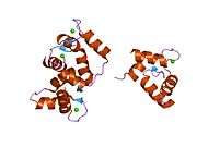
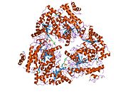
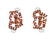
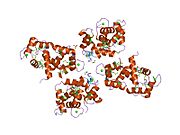
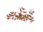
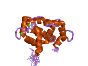
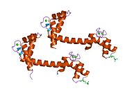
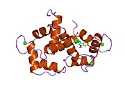
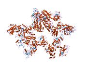
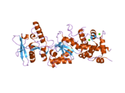
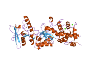
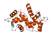
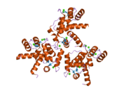
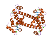
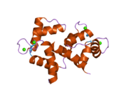
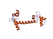
PDB gallery
-
 1a29: CALMODULIN COMPLEXED WITH TRIFLUOPERAZINE (1:2 COMPLEX)
 -
 1ahr: CALMODULIN MUTANT WITH A TWO RESIDUE DELETION IN THE CENTRAL HELIX
 -
 1ak8: NMR SOLUTION STRUCTURE OF CERIUM-LOADED CALMODULIN AMINO-TERMINAL DOMAIN (CE2-TR1C), 23 STRUCTURES
 -
 1cdl: TARGET ENZYME RECOGNITION BY CALMODULIN: 2.4 ANGSTROMS STRUCTURE OF A CALMODULIN-PEPTIDE COMPLEX
 -
 1cdm: MODULATION OF CALMODULIN PLASTICITY IN MOLECULAR RECOGNITION ON THE BASIS OF X-RAY STRUCTURES
 -
 1cfc: CALCIUM-FREE CALMODULIN
 -
 1cfd: CALCIUM-FREE CALMODULIN
 -
 1cff: NMR SOLUTION STRUCTURE OF A COMPLEX OF CALMODULIN WITH A BINDING PEPTIDE OF THE CA2+-PUMP
 -
 1ckk: CALMODULIN/RAT CA2+/CALMODULIN DEPENDENT PROTEIN KINASE FRAGMENT
 -
 1cll: CALMODULIN STRUCTURE REFINED AT 1.7 ANGSTROMS RESOLUTION
 -
 1cm1: MOTIONS OF CALMODULIN-SINGLE-CONFORMER REFINEMENT
 -
 1cm4: MOTIONS OF CALMODULIN-FOUR-CONFORMER REFINEMENT
 -
 1cmf: NMR SOLUTION STRUCTURE OF APO CALMODULIN CARBOXY-TERMINAL DOMAIN
 -
 1cmg: NMR SOLUTION STRUCTURE OF CALCIUM-LOADED CALMODULIN CARBOXY-TERMINAL DOMAIN
 -
 1ctr: DRUG BINDING BY CALMODULIN: CRYSTAL STRUCTURE OF A CALMODULIN-TRIFLUOPERAZINE COMPLEX
 -
 1deg: THE LINKER OF DES-GLU84 CALMODULIN IS BENT AS SEEN IN THE CRYSTAL STRUCTURE
 -
 1dmo: CALMODULIN, NMR, 30 STRUCTURES
 -
 1f70: REFINED SOLUTION STRUCTURE OF CALMODULIN N-TERMINAL DOMAIN
 -
 1f71: REFINED SOLUTION STRUCTURE OF CALMODULIN C-TERMINAL DOMAIN
 -
 1fw4: CRYSTAL STRUCTURE OF E. COLI FRAGMENT TR2C FROM CALMODULIN TO 1.7 A RESOLUTION
 -
 1g4y: 1.60 A CRYSTAL STRUCTURE OF THE GATING DOMAIN FROM SMALL CONDUCTANCE POTASSIUM CHANNEL COMPLEXED WITH CALCIUM-CALMODULIN
 -
 1iq5: Calmodulin/nematode CA2+/Calmodulin dependent kinase kinase fragment
 -
 1iwq: Crystal Structure of MARCKS calmodulin binding domain peptide complexed with Ca2+/Calmodulin
 -
 1j7o: Solution structure of Calcium-calmodulin N-terminal domain
 -
 1j7p: Solution structure of Calcium calmodulin C-terminal domain
 -
 1k90: Crystal structure of the adenylyl cyclase domain of anthrax edema factor (EF) in complex with calmodulin and 3' deoxy-ATP
 -
 1k93: Crystal structure of the adenylyl cyclase domain of anthrax edema factor (EF) in complex with calmodulin
 -
 1l7z: Crystal structure of Ca2+/Calmodulin complexed with myristoylated CAP-23/NAP-22 peptide
 -
 1lin: CALMODULIN COMPLEXED WITH TRIFLUOPERAZINE (1:4 COMPLEX)
 -
 1lvc: Crystal structure of the adenylyl cyclase domain of anthrax edema factor (EF) in complex with calmodulin and 2' deoxy, 3' anthraniloyl ATP
 -
 1mux: SOLUTION NMR STRUCTURE OF CALMODULIN/W-7 COMPLEX: THE BASIS OF DIVERSITY IN MOLECULAR RECOGNITION, 30 STRUCTURES
 -
 1mxe: Structure of the Complex of Calmodulin with the Target Sequence of CaMKI
 -
 1niw: Crystal structure of endothelial nitric oxide synthase peptide bound to calmodulin
 -
 1nwd: Solution Structure of Ca2+/Calmodulin bound to the C-terminal Domain of Petunia Glutamate Decarboxylase
 -
 1ooj: Structural genomics of Caenorhabditis elegans : Calmodulin
 -
 1pk0: Crystal Structure of the EF3-CaM complexed with PMEApp
 -
 1prw: Crystal structure of bovine brain Ca++ calmodulin in a compact form
 -
![1qiv: CALMODULIN COMPLEXED WITH N-(3,3,-DIPHENYLPROPYL)-N'-[1-R-(3,4-BIS-BUTOXYPHENYL)-ETHYL]-PROPYLENEDIAMINE (DPD), 1:2 COMPLEX](//upload.wikimedia.org/wikipedia/commons/thumb/f/f1/PDB_1qiv_EBI.jpg/180px-PDB_1qiv_EBI.jpg) 1qiv: CALMODULIN COMPLEXED WITH N-(3,3,-DIPHENYLPROPYL)-N'-[1-R-(3,4-BIS-BUTOXYPHENYL)-ETHYL]-PROPYLENEDIAMINE (DPD), 1:2 COMPLEX
1qiv: CALMODULIN COMPLEXED WITH N-(3,3,-DIPHENYLPROPYL)-N'-[1-R-(3,4-BIS-BUTOXYPHENYL)-ETHYL]-PROPYLENEDIAMINE (DPD), 1:2 COMPLEX -
![1qiw: CALMODULIN COMPLEXED WITH N-(3,3,-DIPHENYLPROPYL)-N'-[1-R-(3,4-BIS-BUTOXYPHENYL)-ETHYL]-PROPYLENEDIAMINE (DPD)](//upload.wikimedia.org/wikipedia/commons/thumb/4/46/PDB_1qiw_EBI.jpg/180px-PDB_1qiw_EBI.jpg) 1qiw: CALMODULIN COMPLEXED WITH N-(3,3,-DIPHENYLPROPYL)-N'-[1-R-(3,4-BIS-BUTOXYPHENYL)-ETHYL]-PROPYLENEDIAMINE (DPD)
1qiw: CALMODULIN COMPLEXED WITH N-(3,3,-DIPHENYLPROPYL)-N'-[1-R-(3,4-BIS-BUTOXYPHENYL)-ETHYL]-PROPYLENEDIAMINE (DPD) -
 1qs7: The 1.8 angstrom structure of calmodulin rs20 peptide complex
 -
 1qtx: THE 1.65 ANGSTROM STRUCTURE OF CALMODULIN RS20 PEPTIDE COMPLEX
 -
 1qx5: Crystal structure of apoCalmodulin
 -
 1qx7: Crystal structure of apoCaM bound to the gating domain of small conductance Ca2+-activated potassium channel
 -
 1s26: Structure of Anthrax Edema Factor-Calmodulin-alpha,beta-methyleneadenosine 5'-triphosphate Complex Reveals an Alternative Mode of ATP Binding to the Catalytic Site
 -
 1sk6: Crystal structure of the adenylyl cyclase domain of anthrax edema factor (EF) in complex with calmodulin, 3',5' cyclic AMP (cAMP), and pyrophosphate
 -
 1sw8: Solution structure of the N-terminal domain of Human N60D calmodulin refined with paramagnetism based strategy
 -
 1sy9: Structure of calmodulin complexed with a fragment of the olfactory CNG channel
 -
 1up5: CHICKEN CALMODULIN
 -
 1vrk: THE 1.9 ANGSTROM STRUCTURE OF E84K-CALMODULIN RS20 PEPTIDE COMPLEX
 -
 1wrz: Calmodulin complexed with a peptide from a human death-associated protein kinase
 -
 1x02: Solution structure of stereo array isotope labeled (SAIL) calmodulin
 -
 1xa5: Structure of Calmodulin in complex with KAR-2, a bis-indol alkaloid
 -
 1xfu: Crystal structure of anthrax edema factor (EF) truncation mutant, EF-delta 64 in complex with calmodulin
 -
 1xfv: Crystal structure of anthrax edema factor (EF) in complex with calmodulin and 3' deoxy-ATP
 -
 1xfw: Crystal structure of anthrax edema factor (EF) in complex with calmodulin and 3'5' cyclic AMP (cAMP)
 -
 1xfx: Crystal structure of anthrax edema factor (EF) in complex with calmodulin in the presence of 10 millimolar exogenously added calcium chloride
 -
 1xfy: Crystal structure of anthrax edema factor (EF) in complex with calmodulin
 -
 1xfz: Crystal structure of anthrax edema factor (EF) in complex with calmodulin in the presence of 1 millimolar exogenously added calcium chloride
 -
 1y0v: Crystal structure of anthrax edema factor (EF) in complex with calmodulin and pyrophosphate
 -
 1y6w: Trapped intermediate of calmodulin
 -
 1yr5: 1.7-A structure of calmodulin bound to a peptide from DAP kinase
 -
 1yrt: Crystal Structure analysis of the adenylyl cyclaes catalytic domain of adenylyl cyclase toxin of Bordetella pertussis in presence of c-terminal calmodulin
 -
 1yru: Crystal Structure analysis of the adenylyl cyclaes catalytic domain of adenylyl cyclase toxin of Bordetella pertussis in presence of c-terminal calmodulin and 1mM calcium chloride
 -
 1zot: crystal structure analysis of the CyaA/C-Cam with PMEAPP
 -
 1zuz: Calmodulin in complex with a mutant peptide from human DRP-1 kinase
 -
 2bbm: SOLUTION STRUCTURE OF A CALMODULIN-TARGET PEPTIDE COMPLEX BY MULTIDIMENSIONAL NMR
 -
 2bbn: SOLUTION STRUCTURE OF A CALMODULIN-TARGET PEPTIDE COMPLEX BY MULTIDIMENSIONAL NMR
 -
 2bcx: Crystal structure of calmodulin in complex with a ryanodine receptor peptide
 -
 2be6: 2.0 A crystal structure of the CaV1.2 IQ domain-Ca/CaM complex
 -
 2bkh: MYOSIN VI NUCLEOTIDE-FREE (MDINSERT2) CRYSTAL STRUCTURE
 -
 2bki: MYOSIN VI NUCLEOTIDE-FREE (MDINSERT2-IQ) CRYSTAL STRUCTURE
 -
 2col: Crystal structure analysis of CyaA/C-Cam with Pyrophosphate
 -
 2dfs: 3-D structure of Myosin-V inhibited state
 -
 2f2o: Structure of calmodulin bound to a calcineurin peptide: a new way of making an old binding mode
 -
 2f2p: Structure of calmodulin bound to a calcineurin peptide: a new way of making an old binding mode
 -
 2f3y: Calmodulin/IQ domain complex
 -
 2f3z: Calmodulin/IQ-AA domain complex
 -
 2fot: Crystal structure of the complex between calmodulin and alphaII-spectrin
 -
 2hf5: The structure and function of a novel two-site calcium-binding fragment of calmodulin
 -
 2ix7: STRUCTURE OF APO-CALMODULIN BOUND TO UNCONVENTIONAL MYOSIN V
 -
 3cln: STRUCTURE OF CALMODULIN REFINED AT 2.2 ANGSTROMS RESOLUTION
 -
 4cln: STRUCTURE OF A RECOMBINANT CALMODULIN FROM DROSOPHILA MELANOGASTER REFINED AT 2.2-ANGSTROMS RESOLUTION


![1qiv: CALMODULIN COMPLEXED WITH N-(3,3,-DIPHENYLPROPYL)-N'-[1-R-(3,4-BIS-BUTOXYPHENYL)-ETHYL]-PROPYLENEDIAMINE (DPD), 1:2 COMPLEX](http://upload.wikimedia.org/wikipedia/commons/thumb/f/f1/PDB_1qiv_EBI.jpg/180px-PDB_1qiv_EBI.jpg)
![1qiw: CALMODULIN COMPLEXED WITH N-(3,3,-DIPHENYLPROPYL)-N'-[1-R-(3,4-BIS-BUTOXYPHENYL)-ETHYL]-PROPYLENEDIAMINE (DPD)](http://upload.wikimedia.org/wikipedia/commons/thumb/4/46/PDB_1qiw_EBI.jpg/180px-PDB_1qiw_EBI.jpg)


External links
- UniProt.[1] Calm1 Human Gene
- ^ "CALM1 Gene", Definitions, Qeios, 2020-02-07, doi:10.32388/8gddrl, S2CID 243222152, retrieved 2023-10-16


