Aponeurotic fibroma
| Aponeurotic fibroma | |
|---|---|
| Other names | Calcifying aponeurotic fibroma, juvenile aponeurotic fibroma |
 | |
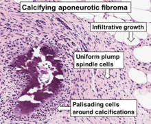
| Histopathology of a calcifying aponeurotic fibroma from a finger, H&E stain. | |
Aponeurotic fibroma, also known as calcifying aponeurotic fibroma, and juvenile aponeurotic fibroma is characterized by a lesion that usually presents as a painless, solitary, deep fibrous nodule, often adherent to tendon, fascia, or periosteum, on the hands and feet.[1] The World Health Organization in 2020 reclassified aponeurotic fibroma nodules as a specific benign type of the fibroblastic and myofibroblastic tumors.[2] Aponeurotic fibromas are diagnosed based on histopathology and treated by surgical excision. They are more common in males than females.
Signs and symptoms
Aponeurotic fibroma occurs most frequently in the fingers, palms, and soles of the distal extremities.[3] Typically, the tumor is defined as a smaller than 3 cm diameter, firm, non-tender mass that grows slowly. It is prone to infiltrate the surrounding tissue and, following surgical resection, is more likely to recur locally.[4]
Diagnosis
A histological examination is necessary to make a diagnosis.[5] Histologically, the tumor is characterized by fibroblast growth and calcification.[3]
Imaging results include edematous alterations and subcutaneous neoplastic tumors with hazy margins that appear to be encroaching on the surrounding tissues. The fascia and tendon sheath are next to the tumor. While T2WI displays heterogeneous signals, T1WI displays signals that are hypointense to isointense. Additionally, there is heterogeneous contrast enhancement.[6][7][8]
Treatment
The treatment of choice for an aponeurotic fibroma is surgical excision.[5]
Epidemiology
Aponeurotic fibroma is a rare tumor. The tumor often manifests in the first or second decade of life, while examples have been documented at birth and 67 years of age. Patients who are male are impacted twice as frequently as those who are female.[4]
See also
- Skin lesion
- Fibromatosis
References
- ^ Freedberg, et al. (2003). Fitzpatrick's Dermatology in General Medicine. (6th ed.). Page 989. McGraw-Hill. ISBN 0-07-138076-0.
- ^ Sbaraglia M, Bellan E, Dei Tos AP (April 2021). "The 2020 WHO Classification of Soft Tissue Tumours: news and perspectives". Pathologica. 113 (2): 70–84. doi:10.32074/1591-951X-213. PMC 8167394. PMID 33179614.
- ^ a b Le, Keasbey (1953). "Juvenile aponeurotic fibroma (calcifying fibroma); a distinctive tumor arising in the palms and soles of young children". Cancer. 6 (2): 338–346. doi:10.1002/1097-0142(195303)6:2<338::aid-cncr2820060218>3.0.co;2-m. ISSN 0008-543X. PMID 13032926. Retrieved 2024-04-17.
- ^ a b Kim, Ok Hwa; Kim, Yeon Mee (2014). "Calcifying Aponeurotic Fibroma: Case Report with Radiographic and MR Features". Korean Journal of Radiology. 15 (1). The Korean Society of Radiology: 134–139. doi:10.3348/kjr.2014.15.1.134. ISSN 1229-6929. PMC 3909846. PMID 24497803.
- ^ a b Sekiguchi, Tomoya; Nakagawa, Motoo; Miwa, Shinji; Shiba, Ayano; Ozawa, Yoshiyuki; Shimohira, Masashi; Sakurai, Keita; Shibamoto, Yuta (2017). "Calcifying aponeurotic fibroma in a girl: MRI findings and their chronological changes". Radiology Case Reports. 12 (3). Elsevier BV: 620–623. doi:10.1016/j.radcr.2017.04.009. ISSN 1930-0433. PMC 5551990.
- ^ Morii, Takeshi; Yoshiyama, Akira; Morioka, Hideo; Anazawa, Ukei; Mochizuki, Kazuo; Yabe, Hiroo (2008). "Clinical significance of magnetic resonance imaging in the preoperative differential diagnosis of calcifying aponeurotic fibroma". Journal of Orthopaedic Science. 13 (3). Elsevier BV: 180–186. doi:10.1007/s00776-008-1226-6. ISSN 0949-2658.
- ^ NISHIO, JUN; INAMITSU, HIDEAKI; IWASAKI, HIROSHI; HAYASHI, HIROYUKI; NAITO, MASATOSHI (2014-07-11). "Calcifying aponeurotic fibroma of the finger in an elderly patient: CT and MRI findings with pathologic correlation". Experimental and Therapeutic Medicine. 8 (3). Spandidos Publications: 841–843. doi:10.3892/etm.2014.1838. ISSN 1792-0981.
- ^ Takaku, Mitsuru; Hashimoto, Ichiro; Nakanishi, Hideki; Kurashiki, Taeko (2011). "Calcifying aponeurotic fibroma of the elbow: a case report". The Journal of Medical Investigation. 58 (1, 2). University of Tokushima Faculty of Medicine: 159–162. doi:10.2152/jmi.58.159. ISSN 1343-1420. PMID 21372502.
Further reading
- Fetsch, John F; Miettinen, Markku (1998). "Calcifying aponeurotic fibroma: A clinicopathologic study of 22 cases arising in uncommon sites". Human Pathology. 29 (12). Elsevier BV: 1504–1510. doi:10.1016/s0046-8177(98)90022-3. ISSN 0046-8177.
- Mocellin, Simone (2021). "Calcifying Aponeurotic Fibroma". Soft Tissue Tumors. Cham: Springer International Publishing. pp. 151–152. doi:10.1007/978-3-030-58710-9_42. ISBN 978-3-030-58709-3.
External links
- DermNet
- Pathology Outlines


